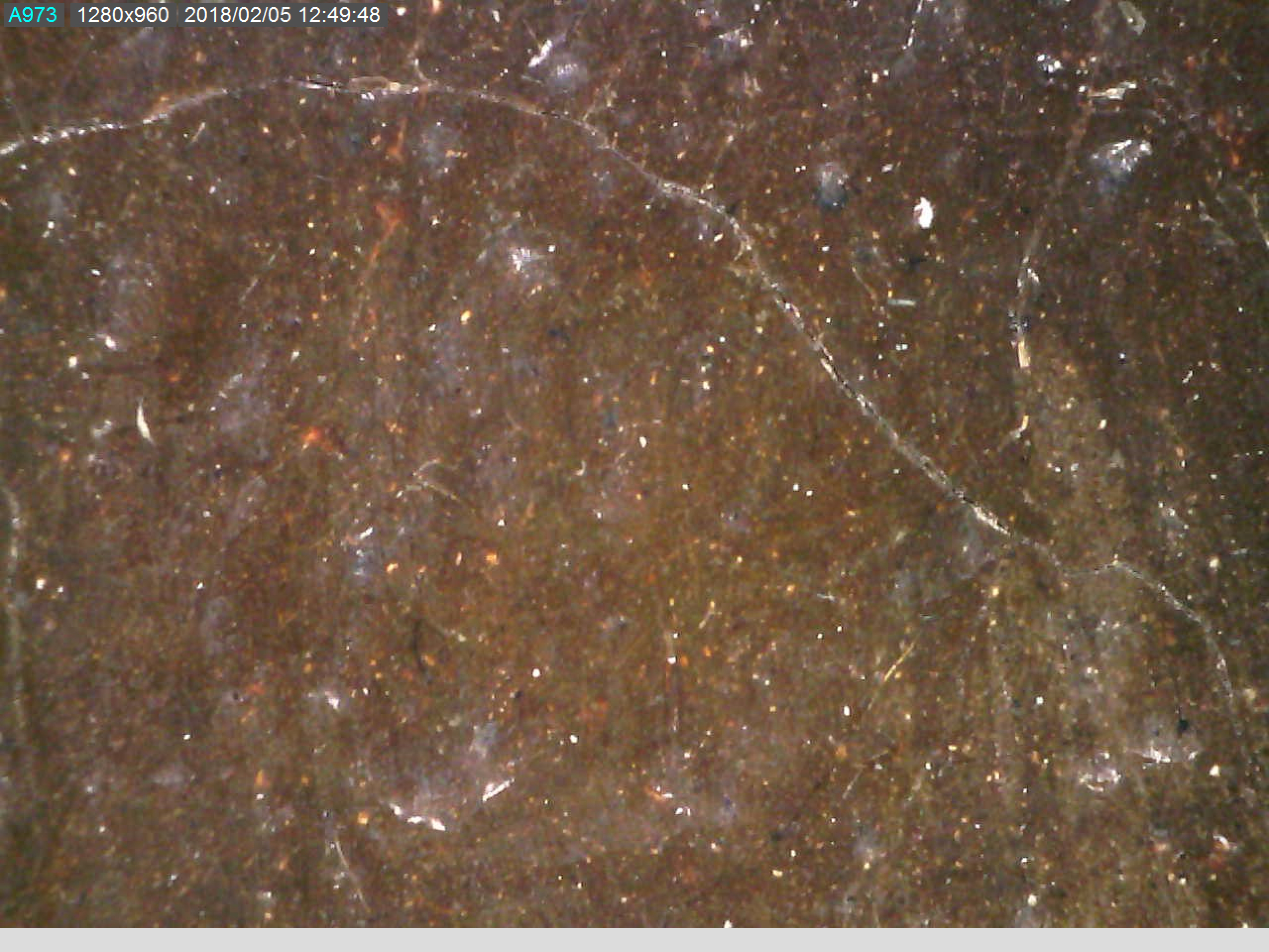
Digital video microscopy is a non-invasive examination technique that lets the surface of the materials that make up the painting, such as the support and the painting layer, to be investigated with magnifications of up to 230x. Video microscopy allows attention to be focused on small sections of the painting, isolating details invisible to the human eye, which cannot distinguish between two points or two lines that are less than 0.2 millimeters apart. It is used in the study of painted surfaces, especially as a diagnostic tool for morphological analysis, and provides thorough information about shape, size and colour. The high magnification power allows us to examine the surfaces affected by different kinds of degradation or restoration interventions, as well as the craquelure, i.e. the dense network of cracks caused by drying or by natural or artificial aging of paint on the pictorial surface. Video microscopy is also used to study the pictorial ductus, i.e. the painter’s brushstrokes, examining their relief, direction, length, curvature, and quality of the strokes and highlighting aspects of the style and features of the artist’s pictorial language. High magnifications analysis, with the aid of X-ray fluorescence spectroscopy, provide to observe the shape, size and colour of the inclusions that make up the backgrounds.